Neuroradiology pays special attention to head, brain, neck and spine health. Your doctor may use an MRI, CT scan or other imaging technologies to detect and treat neurological conditions.
Neuroradiology


An imaging test says a thousand words
Neuroradiology empowers you and your doctor with answers about suspected conditions or how your treatment is progressing. We don’t want you to miss another meaningful moment because of headaches or persistent pain. And when you’ve received a life-changing neurological diagnosis like a brain tumor or multiple sclerosis, we’re here to ease your concerns with personalized treatment plans.
Our neuroradiologists, neurosurgeons and neurologists have devoted their careers to helping people with neurologic conditions live fulfilling lives.
Find a doctor near me

Conditions
Your doctor will turn to neuroradiology to diagnose and treat the following conditions:
Brain tumors
Chronic pain
Epilepsy
Facial pain
Headaches
Movement disorders
Multiple sclerosis
Neurocritical care
Neurodegenerative disorders
Neuromuscular disorders
Stroke
Young adult stroke

Testing
From diagnosing a medical condition to tracking your treatment’s progress, we use MRIs, CT scans and other image-guided procedures as part of your wellness plan.
MRI
Magnetic resonance imaging (MRI) uses a strong magnetic field and radio waves to capture detailed images of the inside of your body. Our team performs the following MRI exams to offer you answers about your health:
- Fetal MRI
- Functional MRI (fMRI)
- MR angiography (MRA)
- Perfusion MRI
Most MRI exams last 20–40 minutes, although compound studies last up to an hour. We’ll get you in and out as quickly as possible so you can safely return to normal life.
CT scans
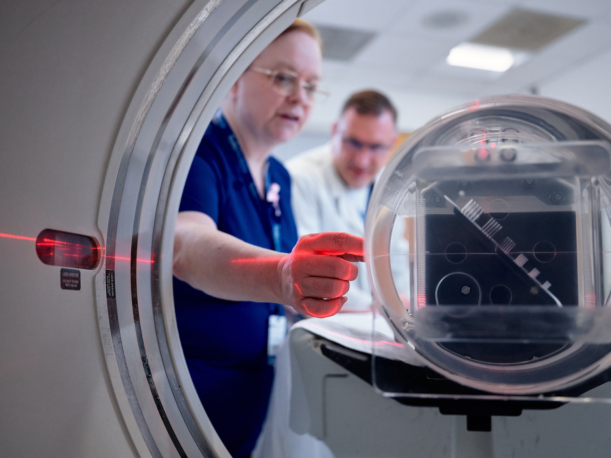
Computerized tomography (CT) scanners combine a series of X-rays into a detailed image of a specific body part, such as your brain, spine, head or neck. Our radiologists can view the scans layer by layer or as a single 3D image.
We offer all forms of CT scans, including:
- CT angiography
- CT perfusion of the head, neck and spine
- CT-guided biopsy
- 3D reconstructions using CT
Image-guided diagnostics
Your doctor may recommend using imaging technology to guide more-involved procedures, such as:
- Lumbar puncture (spinal tap): A needle is inserted into the spinal canal to collect a fluid sample.
- Myelogram: Contrast media is injected into the spinal area to take pictures of your spine and also detect medical conditions.
- CT-guided biopsy: A doctor uses a CT scanner to pinpoint the placement of a needle and draw a tissue sample.
Related services + treatments
Department of Radiology
Academic Department
Our radiologists are helping clinicians see beyond what’s visible through advanced imaging, innovative research and expert education, delivering the diagnostic insights that drive exceptional patient care.